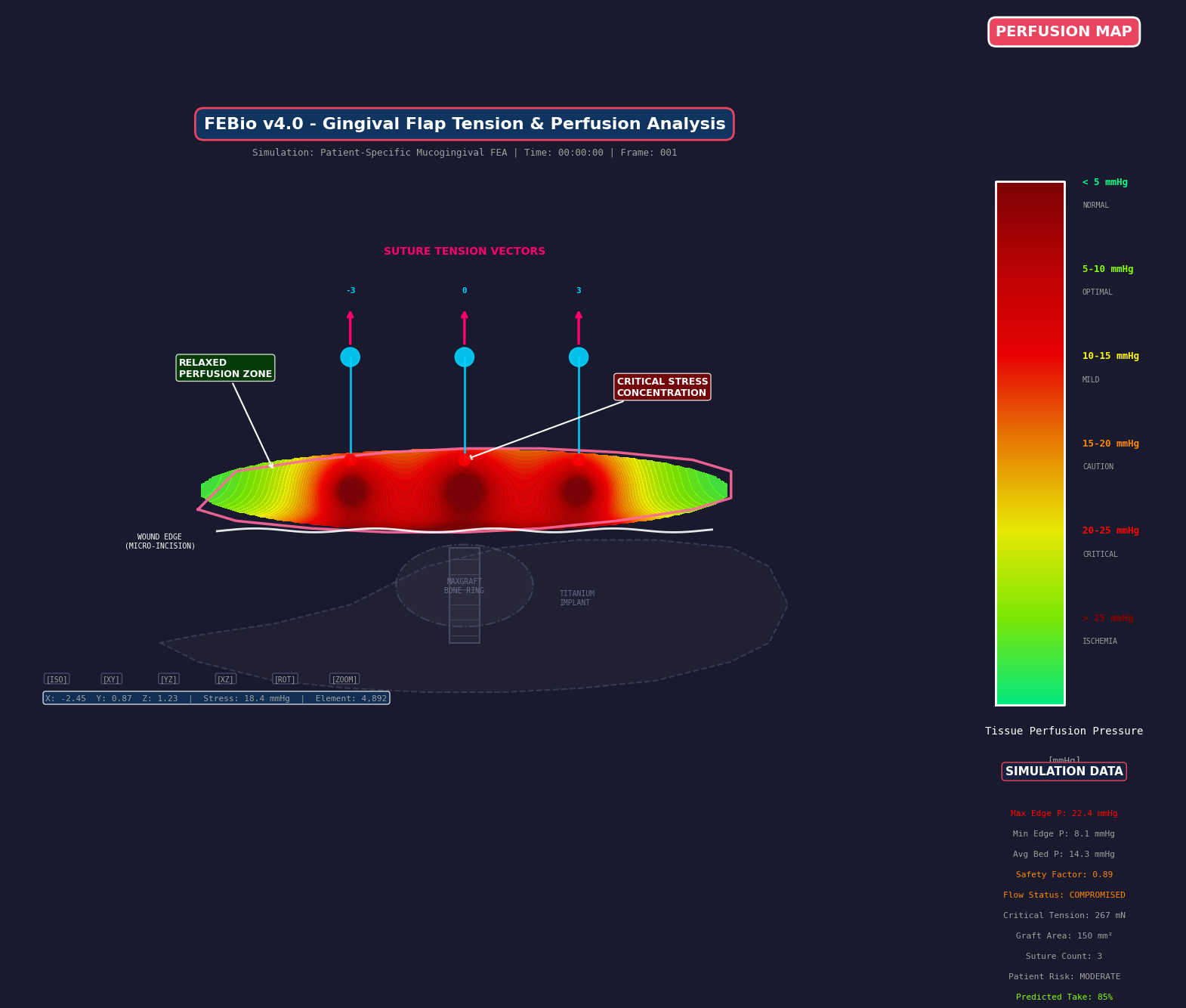
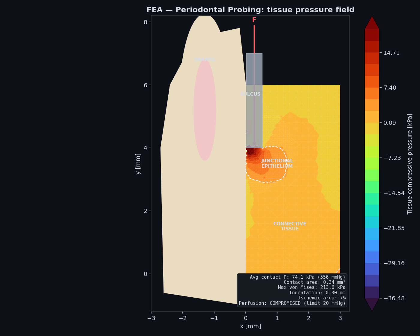
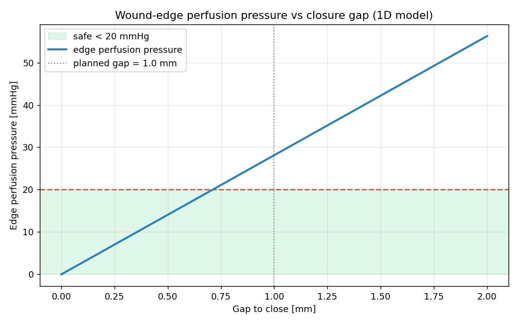
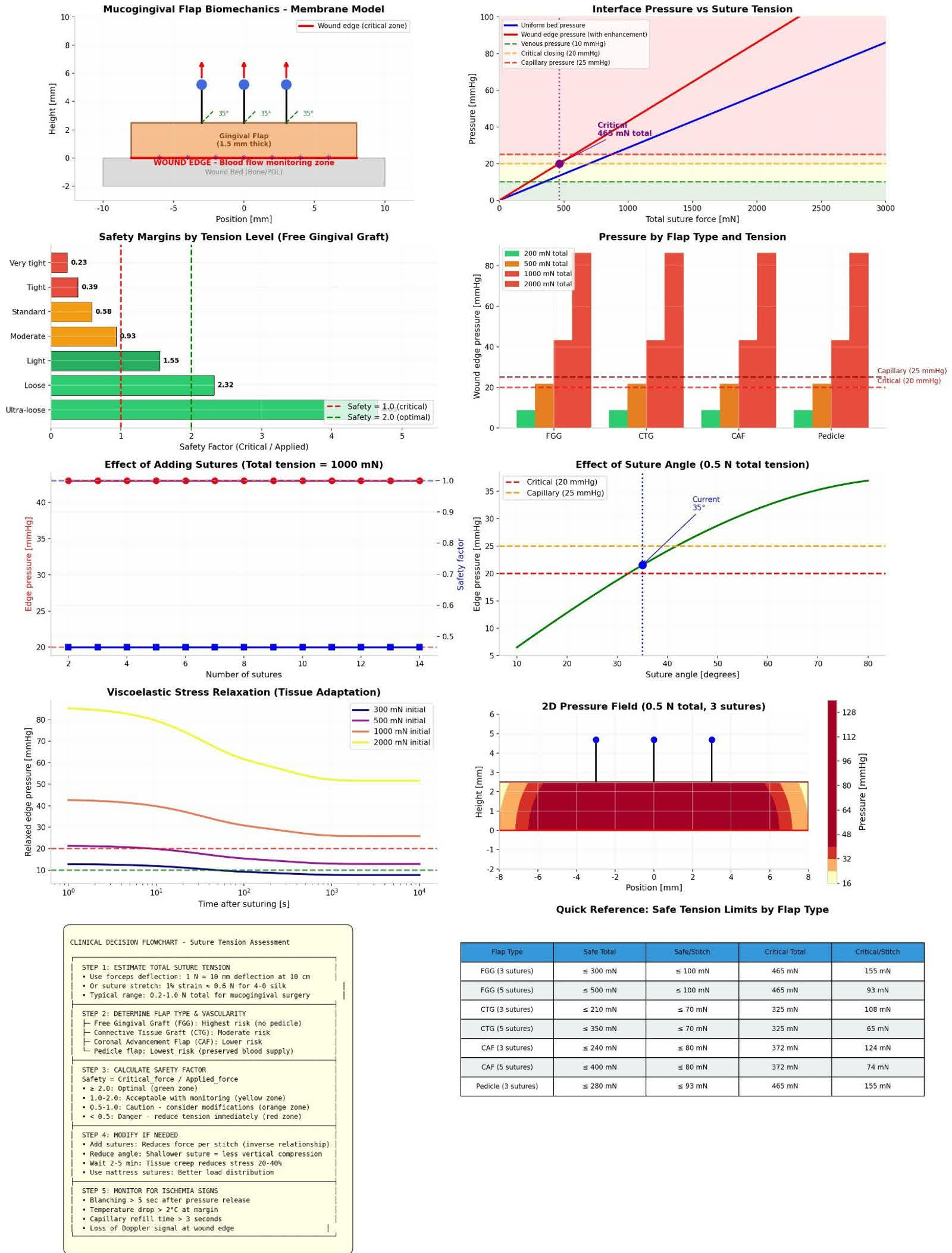
The periodontium is a load-bearing, perfused organ. Planning that ignores walls, perfusion, and force is decorating biology.
Biomechanics, fluid dynamics, perfusion theory, and finite-element analysis — to get into the depths of biology. The plates above are cartoons on purpose. The figures below are teaching models from the same argument. They are not patient-specific diagnosis.
Wall number is a container
A three-wall well is a room. The clot has somewhere to sit. Remaining bone shares load. A two-wall crater still has a chance if you tent the missing face. A one-wall slope is hostile. Zero-wall, suprabony, horizontal loss is a table. The same graft on that table is a different mechanical object. You are not filling a hole. You are stacking on a shelf. Biology cannot pile on a table without a tent.
This is why contained angular defects fill and flat ridges disappoint. Morphology is a boundary condition, not a product choice. The flat-ridge note writes that argument at chair length.
Bone lives near a capillary
Osteocytes are not a powder you sprinkle. They live next to a vessel. A pile on a flat crest is a unilateral healing front: vessels can enter from the bed, not from the denuded root, and not from the oral cavity. Teaching literature puts vascular ingrowth in the neighbourhood of half a millimetre a day. That is a field constant for how far a graft can be asked to wait, not a device output and not a number from my theatre.
Distance from a perfused face is the variable you still control after the harvest. The graft essay names the same fact for soft tissue: it survives on glucose, then on vessels. Bone is not kinder.
PASS is four words, not a kit
Primary closure. Angiogenesis. Space. Stability. Soft-tissue collapse kills the tent. A collagen membrane drapes. It will follow the flap down unless something stiffer holds the volume. Titanium-reinforced membranes tent. They also risk exposure, which is a perfusion event wearing a different name. Urban, Rasperini, and the GTR/GBR field already said this. I am not claiming a trial. I am naming the constraint.
Epithelium always wins the race
Cells do not arrive as a committee. Epithelium is faster than PDL and faster than bone. Guided tissue regeneration is a barrier against that race — a fence so the slow tissues still have a corridor. Enamel matrix derivative is a gel. It can signal. It cannot hold vertical space alone. Asking EMD to reconstruct a horizontal defect is asking a biologic to do a tent's job.
A living fourth wall
When bony walls are gone, a connective-tissue wall can make a living envelope. That is the Rasperini idea I will name as field, not as a percentage. Technique-sensitive. Geometry first. I will not paste a success rate onto a defect I have not seen.
Two organs, one drink
A connective-tissue graft over an avascular root drinks from the flap and the papillae. The root contributes nothing. Volume plus tension is ischemia. A tunnel keeps lateral vessels by not cutting them. A vertical release spends two nutrient faces for a photograph of the defect. Covering a root is covering a dead surface. That sentence belongs on the tray.
Suture tension is perfusion
Blanching is edge pressure crossing capillary closing pressure. Teaching bands sit around 20 mmHg for the colour change. Mechanical dehiscence is a different threshold: Burkhardt and Lang (2010) put a logistic midpoint near 0.15 N. Pini-Prato (2000) already showed that tension costs coverage. Colour can still be acceptable while the suture has left the dehiscence shoulder. They are not the same event. The argument is written: a tight suture is a vascular event. The knobs are on simulate. I am not adding unpublished trial numbers on top.
Recession is a tent on a missing wall
Thin phenotype plus dehiscence dominate. Hard brushing is a mechanical scrape. Plaque inflames. Orthodontics can push a tooth through the bone envelope. After a clean, the swelling leaves and the margin looks lower — that is oedema leaving, not a new disease overnight.
Cairo RT1: no interproximal loss — complete coverage is the expected result. RT2: interproximal loss no worse than buccal — partial coverage is the honest plan. RT3: interproximal worse than buccal — do not sell complete coverage. The house-and-hill picture still holds: soft tissue is a tent; interproximal bone is the pole.
Healing of a free graft is three phases, not a weekend. Roughly day 0–3 plasmatic imbibition; day 2–11 revascularization; day 11–42 maturation, longer if you asked the mucogingival junction to move. Named papers live in the journal. Patient language lives on why gums recede. Chair language for types and walls: look at the papilla first.