Journal · 2026-08-30 · 9 min
A flat ridge is not a cup
Vertical bone on a denuded root with no bony peaks is the hard problem: no container, no lateral blood, and epithelium always wins the race unless you tent and feed.
Igor Kosmina, DMD · Polyclinic Breyer, Sisak · Consultations for a fee
A contained three-wall defect fills because it is a cup. A flat ridge, a denuded root, a zero-wall shelf — that is a table. The same graft on that table is a different mechanical object. We keep pretending it is the same surgery with a different kit.
I work at Polyclinic Breyer in Sisak, and freelance. I do not need a new membrane brand to say this. I need the container named before the product.
The well, and the table
Wall number is a mechanical classification pretending to be an anatomic one. Three walls: a well. The clot has a room. Remaining bone shares load. Two walls: a crater. You can still tent the missing face. One wall: a slope. Hostile. Zero-wall, suprabony, horizontal loss is not a regeneration container. It is a table. Biology cannot pile on a table without a tent.
That sentence is the whole note, if you are in a hurry. The rest is why the tent is so often a rumour.
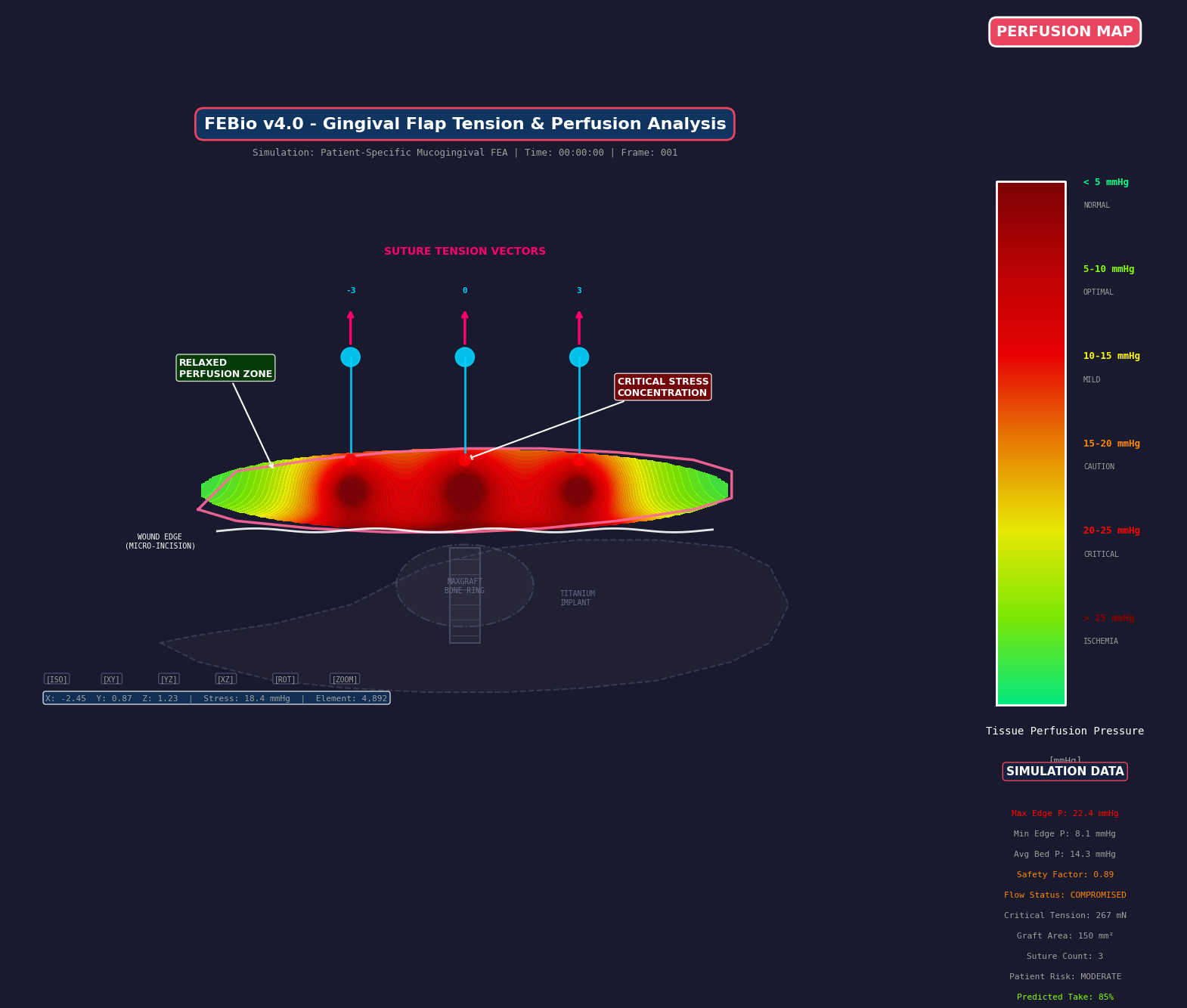
Flap perfusion field. Teaching FEA overlay. Not a medical device. Not for diagnosis.
No lateral blood
Bone lives near a capillary. Osteocytes are not a powder. A pile on a flat crest is a unilateral healing front: vessels can enter from the bed. They cannot enter from a denuded root. They cannot enter from saliva. Teaching literature puts vascular ingrowth in the neighbourhood of half a millimetre a day. That is a field constant for how far a graft can be asked to wait. It is not a device output. It is not a number from my theatre.
A denuded root is an avascular wall. The graft against it drinks from the flap and from whatever papillae you left intact. Volume plus tension is ischemia. Covering that root while also asking for vertical bone is asking one nutrient organ to feed two jobs.
The papilla is the unforgiving cousin. Tarnow et al. (1992): when the contact-to-bone distance is 5 mm or less, the papilla is almost always present. At 6 mm it is there about 56% of the time. At 7 mm, about 27%. Those are published probabilities. They are a field constant, not a promise. If the bony peaks are gone, the black triangle is geometry. You cannot pitch a tent above a missing pole. I already wrote the chair version: look at the papilla first.
PASS is four words
Primary closure. Angiogenesis. Space. Stability. Soft-tissue collapse kills the tent. A collagen membrane drapes. It will follow the flap down unless something stiffer holds the volume. Titanium-reinforced membranes tent. They also risk exposure, which is a perfusion event wearing a different name. The sausage, the tent, the reinforced sheet — those are space-maintenance costumes. They are not a substitute for a wall you do not have.
Urban, the GBR field, the people who actually tent for a living: they already said this. I am naming the constraint in public. I am not pasting a CV percentage on top of their cases.
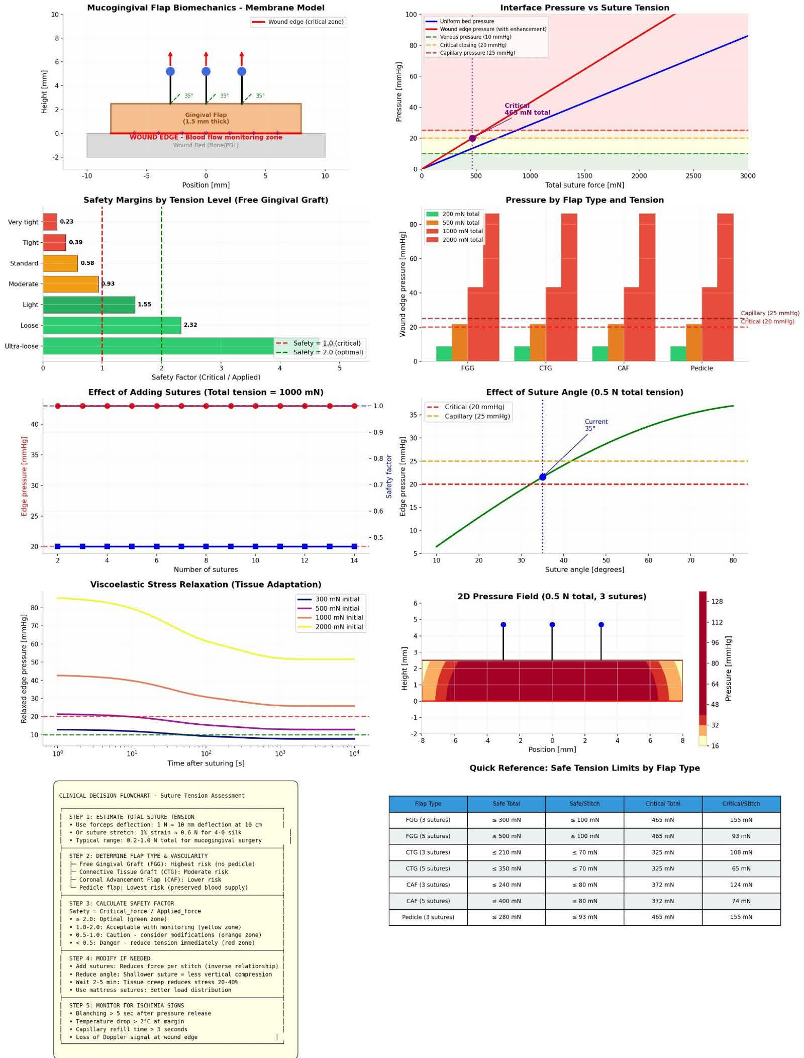
Suture tension and perfusion bands. Teaching dashboard. Not a medical device. Not for diagnosis.
Space without stability is a haematoma with a product name. Stability without primary closure is a membrane in the mouth. Angiogenesis without a bed is a pile. Primary closure with a white flap is a suture that has already spent the capillary.
The race
Cells do not arrive as a committee. Epithelium is faster than PDL. Faster than bone. Guided tissue regeneration is a barrier against that race — a fence so the slow tissues still have a corridor. Remove the fence on a table, and epithelium wins. That is not a moral failure of the patient. It is kinetics.
Enamel matrix derivative is a gel. It can signal. It cannot hold vertical space alone. In a contained well, a biologic has a room to work. In a horizontal defect, asking EMD to reconstruct height is asking a signal to do a tent's job. I will name the biologic as field. I will not pitch it.
A living fourth wall
When bony walls are gone, a connective-tissue wall can make a living envelope. That is the Rasperini idea I will name as field, not as a percentage. A soft-tissue wall is still a wall: it has to be perfused, it has to be stable, it has to not go white under the stitch. Technique-sensitive. Geometry first. I will not paste a 91% onto a defect I have not seen.
Forced eruption belongs in the same paragraph as a named modality, not as a protocol I am handing you. Orthodontic tissue engineering: move the tooth, and sometimes the apparatus comes with it. It is a way of stealing a container from a position the tooth used to occupy. It is not a weekend. It is not this essay's algorithm.
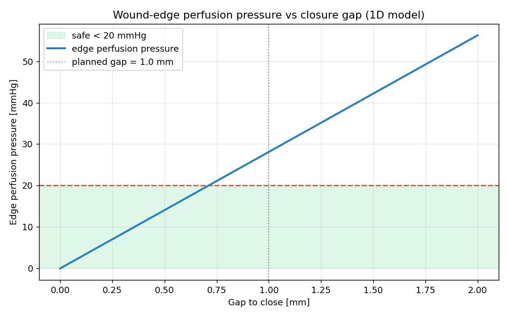
Gap versus edge pressure. Teaching sketch. Not a medical device. Not for diagnosis.
Tension is still perfusion
The flap that tents the table is the same flap I wrote about as a vascular event. Blanching is edge pressure crossing capillary closing pressure. Teaching bands sit around 20 mmHg. Burkhardt and Lang (2010) put mechanical dehiscence near 0.15 N. Pini-Prato (2000) showed tension costs coverage. Colour and dehiscence are not the same event. If you pull the tent white to close a site that had no walls, you have spent the last remaining organ. A tight suture is a vascular event. The knobs are on simulate.
What I am not saying
This is teaching. It is not a surgical algorithm. It is not a certified device output. The FEA plates on this site are overlays on a teaching geometry. They are not patient-specific diagnosis. I am not offering a dual-track clinic menu, a success formula, or a percentage from an unpublished series.
Vertical bone on a denuded root with no bony peaks is the hard problem because there is no container, no lateral blood, and a race epithelium always wins unless you tent and feed. A cup holds. A table spills. If the site is a table, say so before you open. The patient in front of you still needs your chart, not a protocol copied off a slide.
A flat ridge is not a cup.