Patient door
Periodontitis is gum disease that reaches the bone.
Gingivitis is a swollen gum. Periodontitis is that story after the bone has been asked to leave.
Five plates, then a few questions. Pictures first. This is teaching, not a staging of your mouth.
Skip to the stage guideReceding gums?
Educational and illustrative teaching models. Not a medical device. Not for diagnosis, treatment planning, or patient-specific simulation.
The story in five plates
From a tight gum to a tooth that can move
Click a step. The teaching plate and the cross-section move together. Play walks them in order.
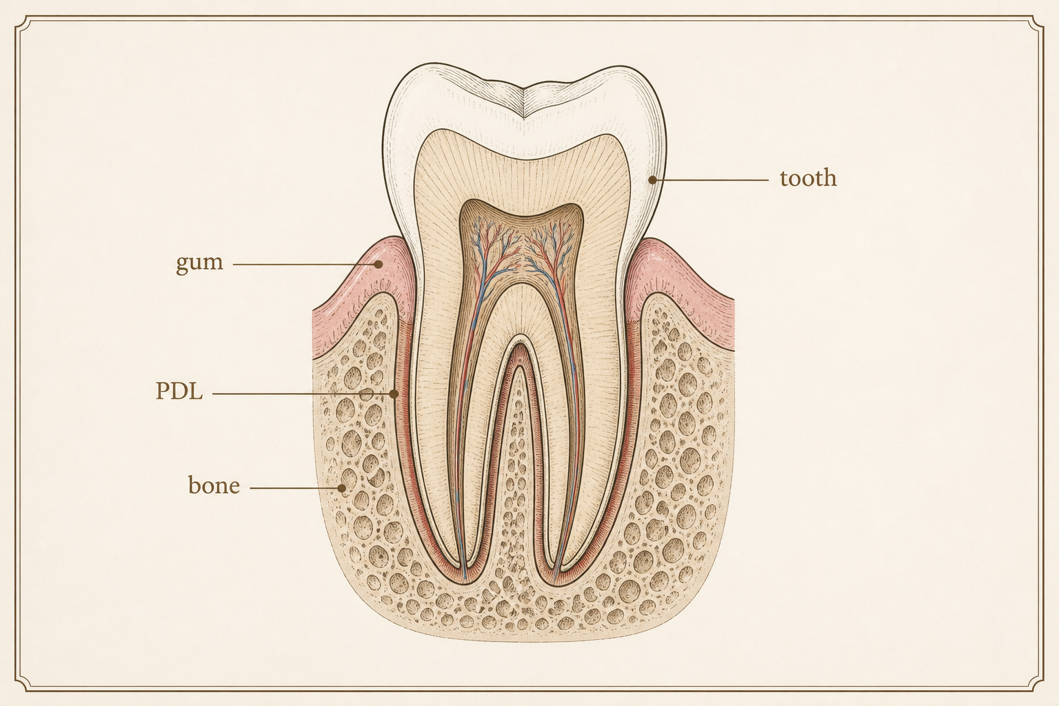
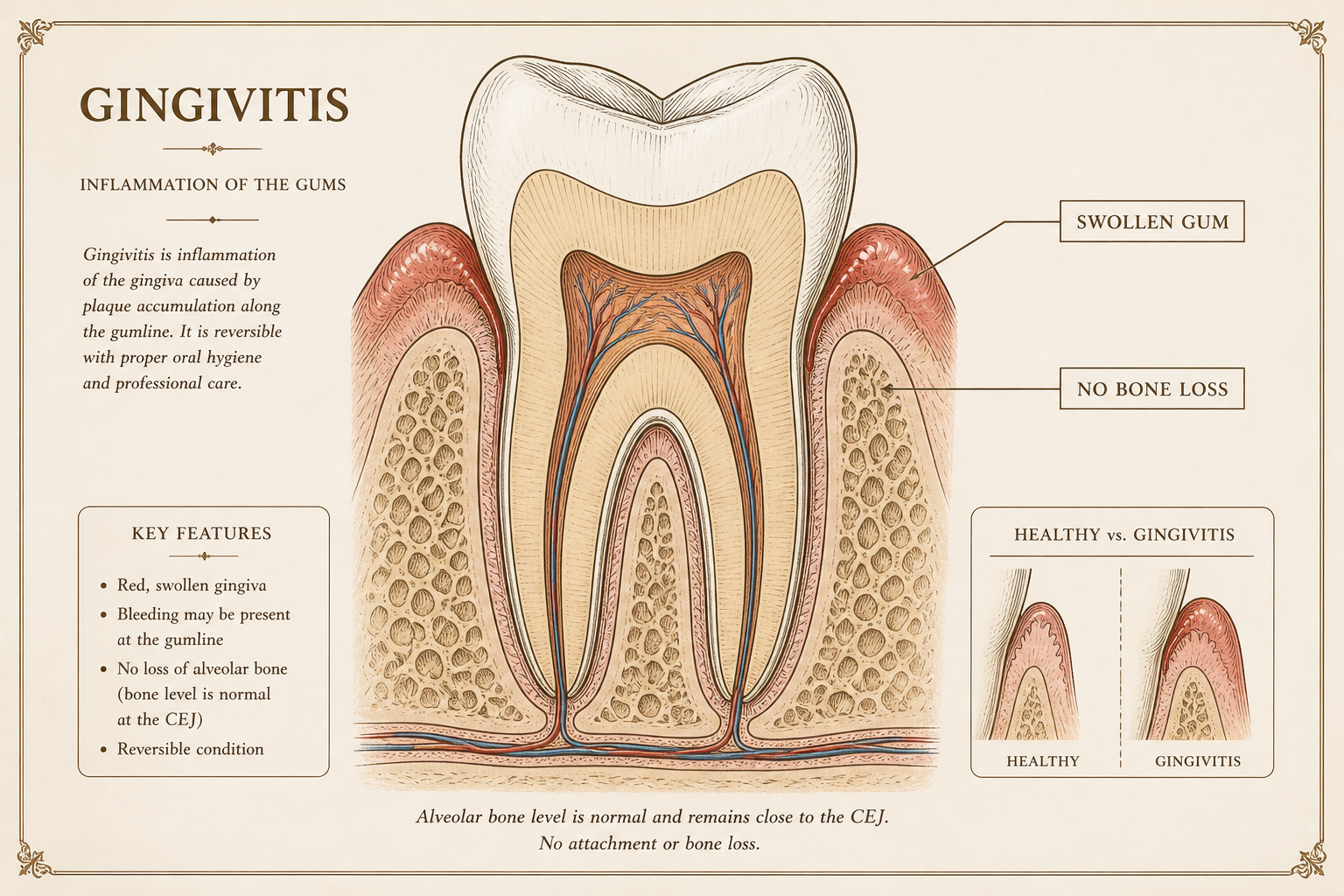
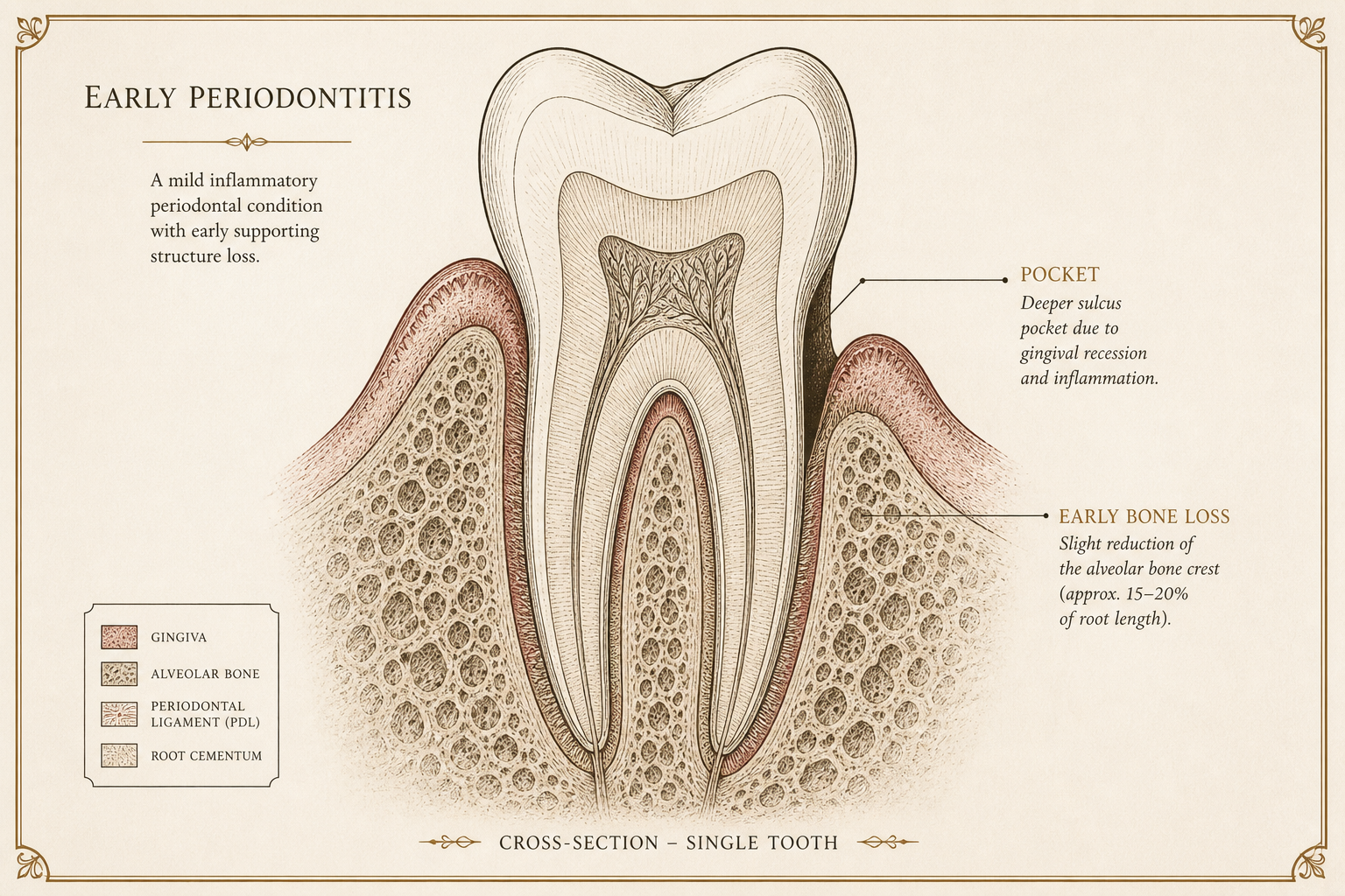
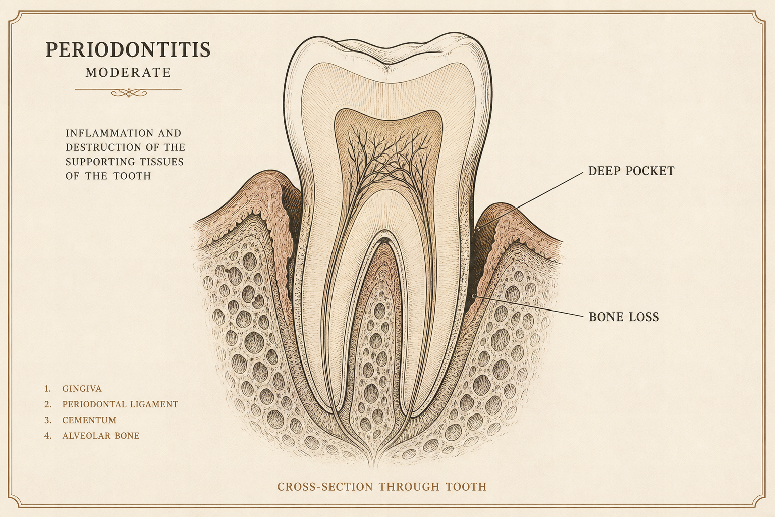
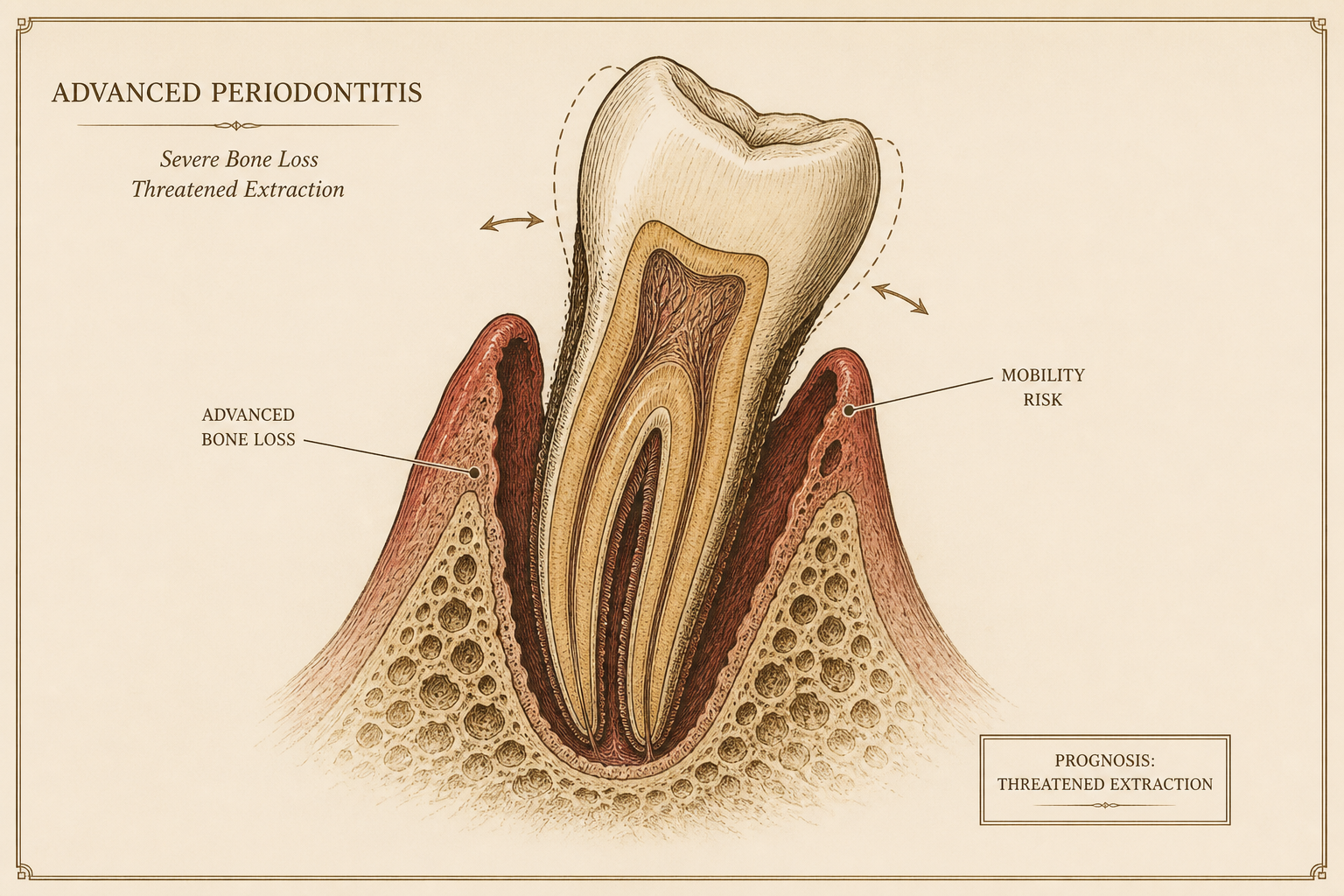
Gums hug the tooth. Bone is high. This is the organ that holds a tooth.
A teaching pattern
Where might this look like you?
Five questions. Not a diagnosis. Answers live in this page until you leave — nothing is stored, no login.
Question 1 of 5
Do your gums bleed when you brush or floss?
If nothing is done
What could happen
Gingivitis can reverse
The gum is allowed to calm. Bone has not left the room yet. That is why we still bother with a clean.
Periodontitis does not grow bone back by itself
Stop the disease and you stop the loss. You do not get a spontaneous refill. Regeneration, when it is honest, is a later, constrained conversation.
Untreated, teeth can loosen
The organ that holds a tooth is bone, ligament, cementum, gum. Take the bone away slowly and the tooth notices.
A destroyed bed is a harder implant
Later implants in a destroyed bed are harder than implants in a socket you still have. That is mechanics, not a scare.
Heart disease and diabetes are linked with periodontitis in research — linked, not a simple cause. It is a reason to take the mouth seriously, not a sentence.
The 2017 staging, if you want the real names
Clinicians, and curious patients. This page does not compute a stage or a grade. Staging needs clinical attachment, radiographs, tooth loss to periodontitis, and complexity. Grading needs rate, smoking, diabetes. Five yes/no answers are not that examination.
Stage I–IV — how much is lost
- I. CAL 1–2 mm. Radiographic bone loss under 15%. No tooth loss to periodontitis. Probing mostly ≤4 mm. Mostly horizontal.
- II. CAL 3–4 mm. Bone loss 15–33%. Still no tooth loss to periodontitis. Probing mostly ≤5 mm.
- III. CAL ≥5 mm. Bone into the middle third of the root and beyond. Up to four teeth lost to periodontitis. Probing ≥6 mm, vertical defects, furcation II/III, moderate ridge defect.
- IV. The same attachment and bone as III, plus five or more teeth lost to periodontitis, or complexity: bite collapse, secondary occlusal trauma, fewer than twenty remaining teeth, a severe ridge defect, a mouth that needs rehabilitation — not only pockets.
Grade A/B/C — how fast
- A. Slow. No loss over five years, or bone loss/age is low. Heavy biofilm, little destruction. Non-smoker. No diabetes.
- B. Moderate. Less than 2 mm over five years. The default if you have not proven A or C.
- C. Rapid. 2 mm or more over five years, or destruction that outruns the biofilm. Smoking ten or more a day, or diabetes with HbA1c ≥7.0, push the grade up.
Direct evidence is change in CAL or bone over time. Indirect is % bone loss divided by age. This walkthrough does not collect either.
If you wanted the chair language for walls and recession types, that lives in the journal: Look at the papilla first. Then count the walls. The ordered plates are in the lab.
Igor Kosmina, DMD · Polyclinic Breyer, Sisak
Also
Receding gums?
A receding gum is a thin tent on a missing wall. Different page. Same organ.
If this looks like a conversation
Write. I am at Polyclinic Breyer in Sisak. Consultations for a fee. The form opens a mail draft — nothing is stored here.
Educational and illustrative teaching models. Not a medical device. Not for diagnosis, treatment planning, or patient-specific simulation.