Journal · 2026-08-31 · 8 min
Mucogingival surgery is geometry
Root coverage is a tent, a wall, and a blood supply. Cairo's recession types are a wall count. Tension is a perfusion event wearing a knot.
Igor Kosmina, DMD · Polyclinic Breyer, Sisak · Consultations for a fee
Do not operate on inflamed tissue. I will start there because the rest of this note is wasted if you skip it.
I work at Polyclinic Breyer in Sisak, and freelance. I do not need a new flap brand. I need the geometry named before the photograph.
Tent, wall, blood
Soft tissue is a tent. Interproximal bone is the pole. You cannot pitch the tent above the pole. The papilla you wish you had is not a flap design. It is an attachment you already measured.
That sentence is the whole note, if you are in a hurry. The rest is why we keep selling height we did not have on the chart.
Cairo's recession type, not Miller, is the soft-tissue sentence. RT1: no interproximal attachment loss — complete coverage is the expected result, and if you do not get it you should have an explanation that is not biology. RT2: interproximal loss less than or equal to the buccal — partial coverage is the honest plan. RT3: interproximal worse than buccal — do not sell complete coverage. Those are missing walls, written in pink. I already wrote the chair version: look at the papilla first. The patient door is why gums recede.
Tarnow et al. (1992): when the contact-to-bone distance is 5 mm or less, the papilla is almost always present. At 6 mm it is there about 56% of the time. At 7 mm, about 27%. Those are published probabilities. They are a field constant, not a promise. If the bony peaks are gone, the black triangle is geometry.
A connective-tissue wall can make a living envelope when bony walls are gone. That is a geometric argument, not a trial claim I will paste onto a defect I have not seen. The envelope still has to be perfused. It still has to not go white under the stitch.
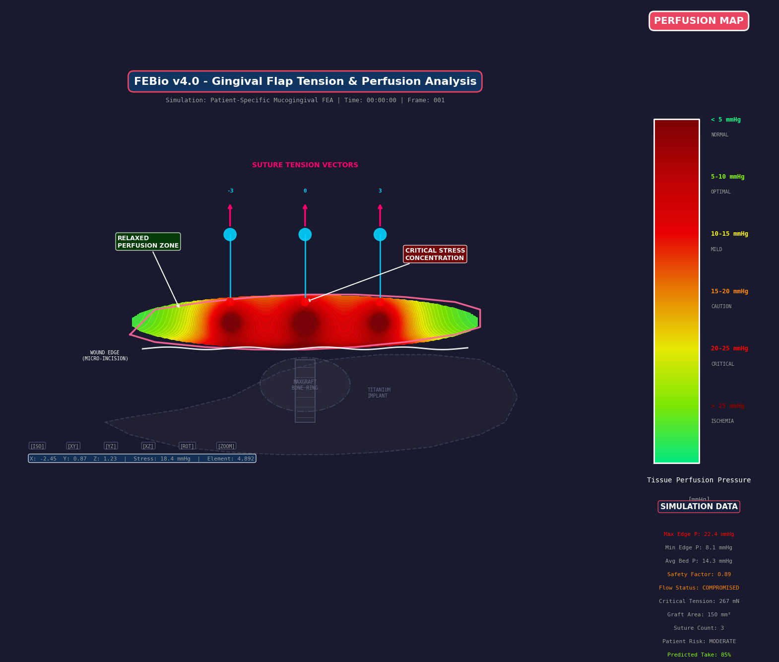
Flap perfusion field. Teaching FEA overlay. Not a medical device. Not for diagnosis.
Tension is still perfusion
The flap that covers the root is a nutrient organ. A denuded root is an avascular wall. The graft against it drinks from the flap and from whatever papillae you left intact. Volume plus tension is ischemia.
Blanching is edge pressure crossing capillary closing pressure. Teaching bands sit around 20 mmHg. Burkhardt and Lang (2010) put mechanical dehiscence near 0.15 N. Pini-Prato (2000) showed tension costs coverage. Colour and dehiscence are not the same event. If you pull the tent white to close a site that had no poles, you have spent the last remaining organ. A tight suture is a vascular event.
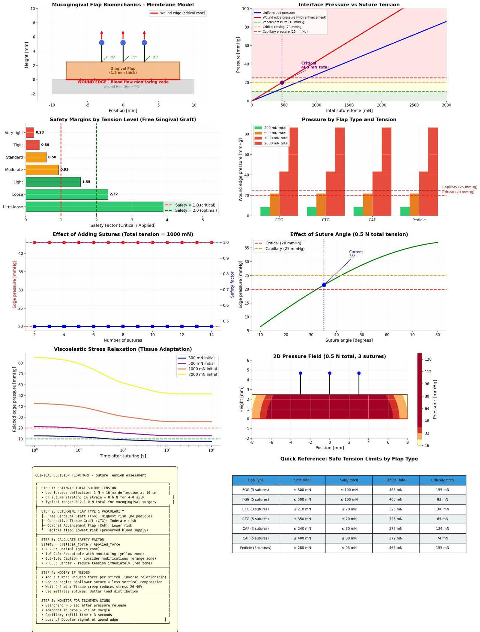
Suture tension and perfusion bands. Teaching dashboard. Not a medical device. Not for diagnosis.
A tunnel preserves lateral supply by skipping vertical releases. That is perfusion again, wearing a different acronym. If you release vertically because the access is prettier, you have spent two nutrient faces for a photograph of the defect. Wound stability, space, tension-free primary closure: those are why the tent you counted still exists at day ten. They are not a product line.
What I am not saying
This is not a surgical algorithm. It is not the best mucogingival surgery. I will not rank tunnels against coronally advanced flaps as if a kit settled geometry. I will not give you a fake referral desk. I will not paste a complete-coverage percentage onto an RT3.
Healing of a free graft is phases, not a weekend. Plasmatic imbibition first. Then vessels. Then maturation. Named papers live in the sibling notes. Distance from a perfused face is still the variable you can change after the harvest: the graft survives on glucose.
This is teaching. The FEA plates are overlays on a teaching geometry. They are not patient-specific diagnosis. The patient in front of you still needs your chart, not a protocol copied off a slide.
If you wanted the ordered plates, the lab is the path. The sibling notes are the chair versions: papilla first, then the stitch. Look at the pole. Count the walls. Then decide whether you are covering a root or filling a well. Those are different surgeries.
Mucogingival surgery is geometry.